Welcome to the Amira-Avizo Software Use Case Gallery
Below you will find a collection of use cases of our 3D data visualization and analysis software. These use cases include scientific publications, articles, papers, posters, presentations or even videos that show how Amira-Avizo Software is used to address various scientific and industrial research topics.
Use the Domain selector to filter by main application area, and use the Search box to enter keywords related to specific topics you are interested in.
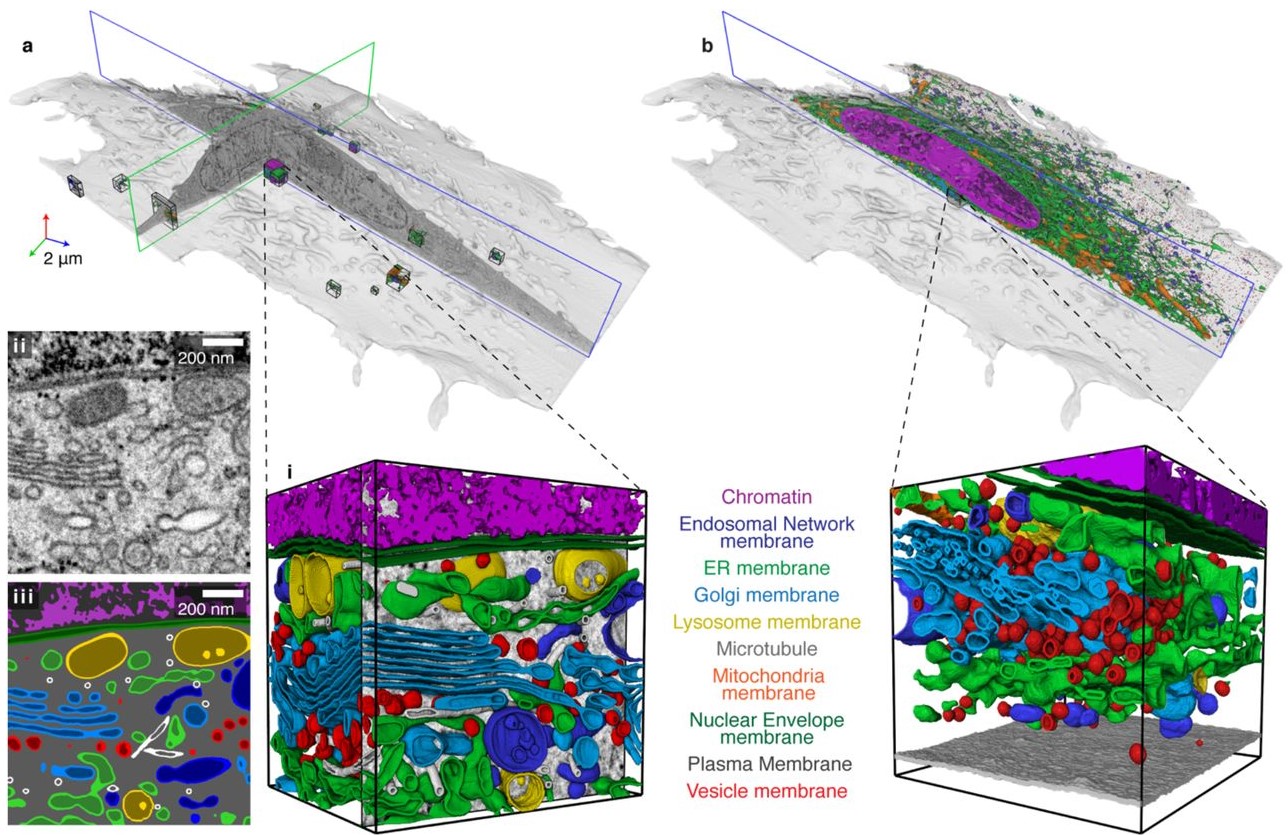
Automatic whole cell organelle segmentation in volumetric electron microscopy
Cells contain hundreds of different organelle and macromolecular assemblies intricately organized relative to each other to meet any cellular demands. Obtaining a complete understanding of their organization is challenging and requires nanometer-level, three-dimensional reconstruction of whole cells. Even then, the immense size of datasets and large number of structures to be characterized requires generalizable, automatic methods.
To meet this challenge, we developed an analy... Read more
Larissa Heinrich, Davis Bennett, David Ackerman, Woohyun Park, John Bogovic, View ORCID ProfileNils Eckstein, Alyson Petruncio, Jody Clements, C. Shan Xu, Jan Funke, Wyatt Korff, Harald F. Hess, Jennifer Lippincott-Schwartz, Stephan Saalfeld, Aubrey V. Weigel, COSEM Project Team
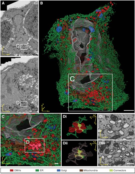
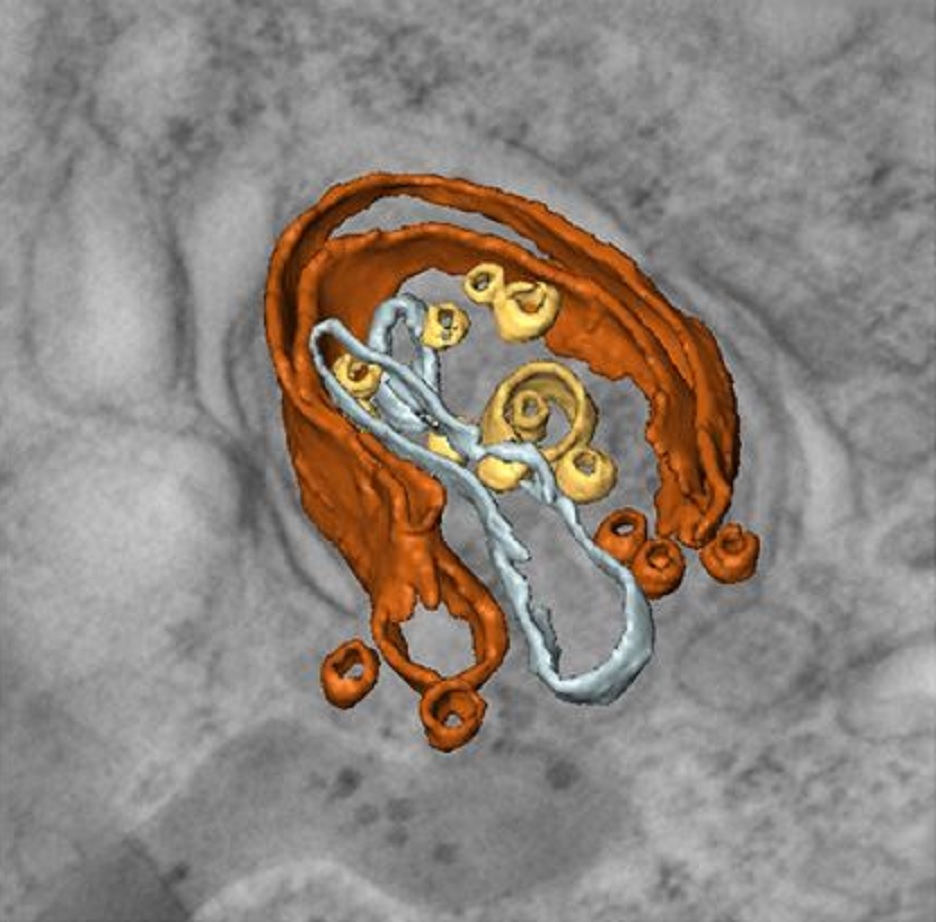
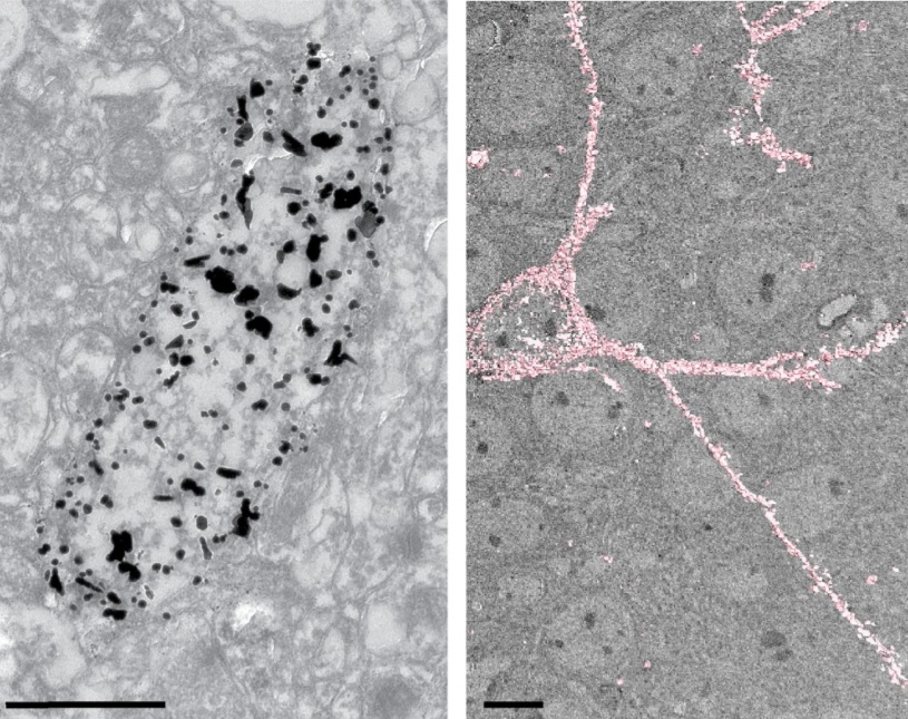
Integrative Imaging Reveals SARS-CoV-2-Induced Reshaping of Subcellular Morphologies
Cortese et al. use integrative imaging techniques to generate a publicly available repository of morphological alterations induced by SARS-CoV-2 in lung cells. Accumulation of ER-derived double-membrane vesicles, the viral replication organelle, occurs concomitantly with cytoskeleton remodeling and Golgi fragmentation. Pharmacological alteration of cytoskeleton dynamics restricts viral replication and spread.
Pathogenesis induced by SARS-CoV-2 is thought to result from both an inflamma... Read more
Mirko Cortese, Ji-Young Lee, Berati Cerikan, ..., Laurent Chatel-Chaix, Yannick Schwab, Ralf Bartenschlager
Macroautophagy is morphologically characterized by autophagosome formation. Autophagosomes are double-membraned vesicles that sequester cytoplasmic components for further degradation in the lysosome. Basal autophagy is paramount for intracellular quality control in post-mitotic cells but, surprisingly, the number of autophagosomes in post-mitotic neurons is very low, suggesting that alternative degradative structures could exist in neurons…
Read more
Maria Rosario Fernandez-Fernandez, Desire Ruiz-Garcia, Eva Martin-Solana, Francisco Javier Chichon, Jose L. Carrascosa, Jose-Jesus Fernandez
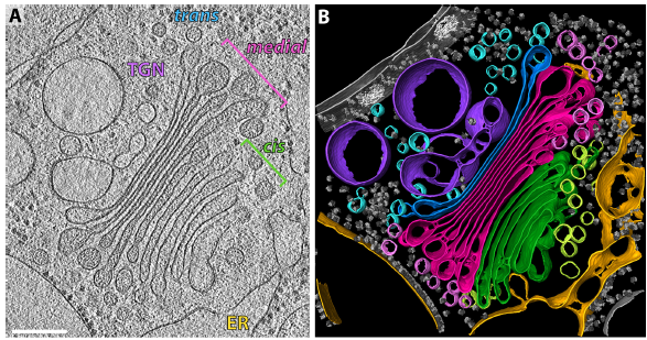
The structure of the COPI coat determined within the cell
COPI-coated vesicles mediate trafficking within the Golgi apparatus and from the Golgi to the endoplasmic reticulum. Here, we applied cryo-focused ion beam milling, cryo-electron tomography and subtomogram averaging to determine the native structure of the COPI coat within vitrified Chlamydomonas reinhardtii cells. The native algal structure resembles the in vitro mammalian structure, but additionally reveals cargo bound beneath beta’–COP. We find that all coat components disassemble... Read more
Yury S Bykov, Miroslava Schaffer, Svetlana O Dodonova, Sahradha Albert, Jurgen M Plitzko, Wolfgang Baumeister, Benjamin D Engel, John AG Briggs
Analysis of neuronal arborization and connections is a powerful tool in fundamental and clinical neuroscience. Changes in neuronal morphology are central to brain development and plasticity and are associated with numerous diseases. Golgi staining is a classical technique based on a deposition of metal precipitate in a random set of neurons. Despite their versatility, Golgi methods have limitations that largely precluded their use in advanced microscopy. We combined Golgi staining with fluore... Read more
Katlijn Vints, Dorien Vandael, Pieter Baatsen, Benjamin Pavie, Frank Vernaillen, Nikky Corthout, Vasily Rybakin, Sebastian Munck & Natalia V. Gounko
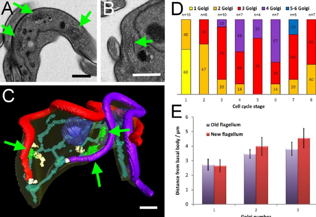
The major mammalian bloodstream form of the African sleeping sickness parasite Trypanosoma bruceimultiplies rapidly, and it is important to understand how these cells divide. Organelle inheritance involves complex spatiotemporal re-arrangements to ensure correct distribution to daughter cells…
Read more
Louise Hughes, Samantha Borrett, Katie Towers, Tobias Starborg, Sue Vaughan
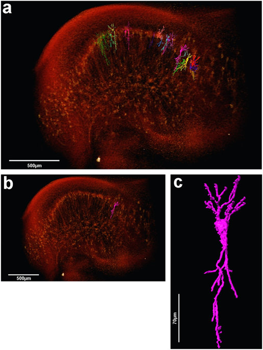
The assessment of neuronal number, spatial organization and connectivity is fundamental for a complete understanding of brain function. However, the evaluation of the three-dimensional (3D) brain cytoarchitecture at cellular resolution persists as a great challenge in the field of neuroscience. In this context, X-ray microtomography has shown to be a valuable non-destructive tool for imaging a broad range of samples, from dense materials to soft biological specimens, arisen as a new method fo... Read more
Matheus de Castro Fonseca, Bruno Henrique Silva Araujo, Carlos Sato Baraldi Dias, Nathaly Lopes Archilha, Dionísio Pedro Amorim Neto, Esper Cavalheiro, Harry Westfahl Jr, Antônio José Roque da Silva, Kleber Gomes Franchini